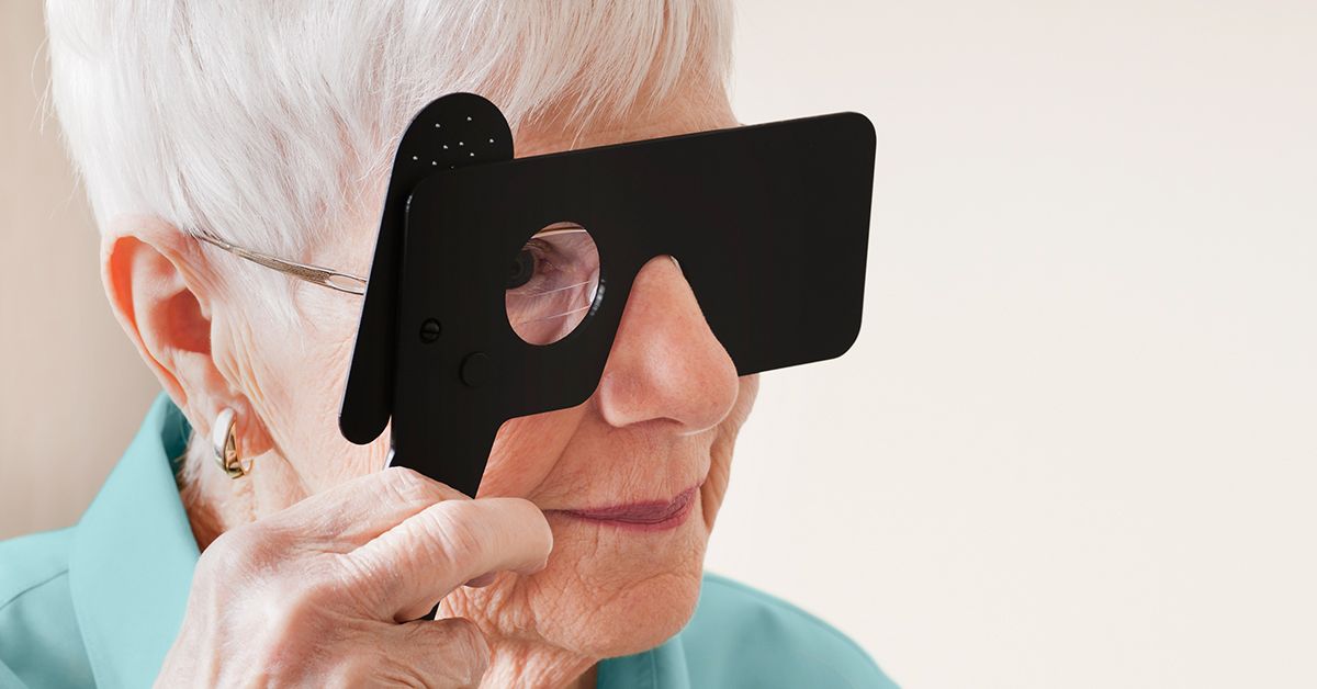
When blurry vision sends you to an eye doctor, you may expect high-tech cameras, glowing screens, and equipment that looks ready to scan a distant planet. Instead, the doctor may begin by asking you to look through a tiny hole in a simple handheld card.
That surprisingly modest procedure is called the pinhole test. It helps an eye care professional estimate whether reduced visual acuity is mainly caused by a refractive error, such as nearsightedness or astigmatism, or whether an eye condition may be limiting vision. The test is quick, painless, inexpensive, and remarkably useful for something that resembles a very serious piece of Swiss cheese.
However, the pinhole test does not diagnose age-related macular degeneration, commonly called AMD. It provides a clue. If vision remains blurry through the pinhole, the doctor may investigate conditions involving the retina, macula, lens, cornea, optic nerve, or other parts of the visual system. A comprehensive dilated eye examination and retinal imaging are needed to confirm macular degeneration.
What Is the Pinhole Eye Test?
The pinhole test is a basic visual acuity assessment performed with an opaque occluder containing one or more tiny openings. During the test, the patient covers one eye, looks through a pinhole with the other eye, and reads letters or symbols on a standard eye chart.
The clinician compares two measurements:
- Visual acuity without the pinhole
- Visual acuity while looking through the pinhole
A meaningful improvement usually indicates that at least part of the blur is related to an uncorrected or undercorrected refractive error. In plain English, the eye may need a better glasses or contact lens prescription.
If vision improves little or not at all, the problem may not be correctable with ordinary lenses alone. The doctor may then look for cataracts, corneal disease, retinal disorders, macular degeneration, optic nerve damage, or another cause of reduced vision. The American Academy of Ophthalmology and American Optometric Association include pinhole acuity among the useful components of visual assessment when clinically indicated.
How Does the Pinhole Test Work?
To understand the test, imagine sunlight passing through a small gap in a window shade. The narrow opening limits the number of light rays entering the room. A pinhole occluder does something similar for the eye.
Normally, the cornea and lens bend incoming light so it focuses precisely on the retina. With a refractive error, some rays focus in front of, behind, or unevenly across the retina. The result is a blurred image.
The pinhole blocks many of the unfocused peripheral light rays and allows a narrower central bundle to reach the retina. This reduces the size of the blur circle and temporarily increases depth of focus. The test does not repair the eye, change the prescription, or treat a disease. It simply creates a controlled optical shortcut.
This explains why someone with an outdated glasses prescription may suddenly read extra letters through the pinhole. The improvement suggests that proper refraction could sharpen vision. It is essentially the eye-care version of saying, “The camera may be fine; let us adjust the focus.”
What Does the Pinhole Test Have to Do With Macular Degeneration?
The macula is the small central area of the retina responsible for detailed straight-ahead vision. It helps with reading, recognizing faces, driving, seeing colors, and performing tasks that require fine detail. Macular degeneration damages this central retinal tissue, so the retina may be unable to process a perfectly focused image normally.
Because a pinhole mainly corrects optical defocus, it cannot restore retinal cells damaged by AMD. A patient whose poor central vision is primarily caused by macular degeneration may therefore show limited improvement on the pinhole test.
For example, suppose a patient reads only the 20/80 line without correction:
- If the patient reaches 20/30 through the pinhole, refractive error probably accounts for much of the blur.
- If the patient remains around 20/80, an eye condition may be limiting visual potential.
- If the patient improves to 20/50, both refractive error and underlying eye disease may be contributing.
These examples illustrate possibilities, not rigid diagnostic rules. Pinhole performance varies with the severity and location of retinal damage, lighting, pupil size, cataracts, the patient’s ability to align with the opening, and the accuracy of the original visual acuity measurement.
Can the Pinhole Test Diagnose Macular Degeneration?
No. The pinhole test cannot confirm or rule out macular degeneration.
A poor response may raise concern about ocular disease, but it does not identify which disease is present. Cataracts, corneal scarring, diabetic retinopathy, macular holes, retinal detachment, optic nerve disease, and several other conditions can also prevent pinhole improvement.
Likewise, some people with early AMD may still have relatively good chart vision or may improve through a pinhole because they also have an uncorrected refractive error. Early and intermediate AMD can exist before a person notices significant symptoms, which is one reason routine dilated examinations are so important. The National Eye Institute emphasizes that a comprehensive dilated eye exam is needed to detect AMD, particularly in its earlier stages.
How the Pinhole Test Is Performed
1. Your regular visual acuity is measured
You will usually read a Snellen chart or another standardized eye chart from a set distance. One eye is covered while the other is tested. The smallest line read accurately becomes the baseline measurement.
2. The pinhole occluder is positioned
The clinician places the occluder in front of the tested eye. If it contains several openings, you may be asked to move it slightly until you find the clearest view.
3. You read the chart again
You attempt the same chart while looking only through the small opening. The clinician records whether you can identify more letters or reach a smaller line.
4. The results are compared
Improvement of one line may be noted, but greater improvement generally provides stronger evidence that refractive error is contributing. The clinician interprets the change alongside the rest of the examination rather than treating it as a mathematical verdict from the Eye Court of Appeals.
The procedure usually takes only a minute or two. It requires no eye drops, injections, radiation, or contact with the surface of the eye.
How Doctors Interpret Pinhole Test Results
Vision improves substantially
A strong improvement usually points toward myopia, hyperopia, astigmatism, or an outdated prescription. A formal refraction can then determine the lens power needed for the best possible correction. Merck Manual guidance describes pinhole improvement as a rapid way to identify blur likely caused by refractive error.
Vision does not improve
No improvement suggests that simply focusing light more accurately may not solve the problem. The doctor may evaluate the cornea, lens, vitreous, retina, macula, optic nerve, and visual pathways.
In an older adult with central distortion, faded colors, difficulty recognizing faces, or a dark spot in the center of vision, poor pinhole improvement can support the need for a detailed macular evaluation. It still does not prove that AMD is responsible.
Vision improves only partially
Partial improvement is common when more than one issue is present. A patient might have astigmatism plus cataracts, or an outdated prescription plus macular degeneration. Correcting the refractive component may improve day-to-day vision even though an underlying retinal condition remains.
Vision becomes worse through the pinhole
A smaller aperture reduces the amount of light reaching the retina. People with dense cataracts, central lens opacities, macular disease, poor contrast sensitivity, or difficulty aligning with the opening may perform worse. Research also shows that pinhole acuity does not perfectly predict best-corrected vision in every patient, particularly when retinal pathology is present.
Which Tests Actually Help Diagnose Macular Degeneration?
When AMD is suspected, the pinhole test is only one small piece of a much larger puzzle. The following assessments provide more direct information about the macula.
Dilated retinal examination
Dilating drops enlarge the pupils so the eye doctor can examine the retina and optic nerve. The doctor looks for drusen, pigment changes, bleeding, fluid, geographic atrophy, scarring, and abnormal blood vessels.
Amsler grid
The Amsler grid is often confused with the pinhole test, but the two serve different purposes. The grid consists of straight horizontal and vertical lines with a central dot. A patient with macular changes may notice lines that appear wavy, missing, dim, distorted, or broken.
The grid can help monitor central vision at home, especially in people already diagnosed with AMD. A new distortion should be reported promptly rather than saved for the next routine appointment.
Optical coherence tomography
Optical coherence tomography, or OCT, uses light waves to create detailed cross-sectional images of the retina. It can reveal drusen, retinal thinning, fluid, swelling, pigment epithelial changes, and other structural abnormalities associated with dry or wet AMD.
OCT is painless and noninvasive. Unlike the pinhole test, which gives an indirect clue about visual blur, OCT lets the clinician inspect the retinal layers themselves. It has become an essential tool for diagnosing and monitoring macular disease.
Fundus photography
Retinal photographs document the appearance of the macula and allow changes to be compared over time. They can record drusen, pigmentation abnormalities, hemorrhage, or atrophy.
Fluorescein angiography
When wet AMD is suspected, a fluorescent dye may be injected into a vein while a specialized camera photographs circulation through the retina. The images can show leaking abnormal blood vessels.
OCT angiography
OCT angiography can map retinal and choroidal blood flow without injected dye. It may help detect or monitor abnormal blood vessel networks, although the clinician decides which imaging method is appropriate for each case.
Pinhole Test Versus Amsler Grid
The names are sometimes mentioned together during an eye examination, but they answer different questions.
| Feature | Pinhole Test | Amsler Grid |
|---|---|---|
| Main purpose | Estimates whether blur may improve with refractive correction | Checks for distortion or missing areas in central vision |
| Common setting | Eye clinic, medical office, or vision screening | Eye clinic and home monitoring |
| Typical target | Letters or symbols on an eye chart | A square grid with a central dot |
| Can it diagnose AMD? | No | No, but abnormal results can signal macular changes |
| What an abnormal result suggests | Reduced vision may not be caused solely by refractive error | Central retinal function may have changed |
Limitations of the Pinhole Test
Although useful, the pinhole test has several important limitations:
- It does not identify the exact cause of reduced vision.
- It cannot show drusen, bleeding, retinal fluid, or macular atrophy.
- It may underestimate potential vision when the opening is poorly aligned.
- Reduced illumination can make the chart harder to see.
- Older adults may have difficulty holding their head and eye in the correct position.
- Very large refractive errors may not fully correct through a standard pinhole.
- Cataracts and retinal disease may produce mixed or misleading responses.
- Good pinhole acuity does not exclude early macular degeneration.
For these reasons, eye care professionals interpret the test as a screening clue, not a final diagnosis or substitute for refraction, dilation, retinal examination, or imaging.
When Should Changes in Central Vision Be Evaluated?
Arrange a prompt eye examination when you notice persistent blurring, trouble reading despite adequate lighting, difficulty recognizing faces, reduced color brightness, or a new central blind spot.
Seek urgent eye care for sudden distortion, rapidly worsening central vision, new dark or blank areas, flashes of light, a sudden increase in floaters, or a curtain-like shadow. These symptoms may indicate wet AMD or another retinal emergency. Waiting to see whether the problem “settles down” can sacrifice valuable treatment time.
A pinhole device purchased online should not be used to diagnose yourself. Temporary clarity through a tiny opening does not prove that your eyes are healthy, and a poor response does not prove that you have macular degeneration.
Experiences With the Pinhole Test and Macular Degeneration
The following composite scenarios reflect experiences commonly discussed in eye-care settings. They are educational examples rather than accounts of specific patients.
The surprisingly clearer eye chart
One person arrives convinced that macular degeneration has suddenly become worse because street signs look fuzzy and television captions are harder to read. Without glasses, the eye chart is a foggy collection of black shapes. Through the pinhole, several smaller letters become visible.
That moment can feel encouraging, but it does not erase the AMD diagnosis. It suggests that an outdated prescription is adding avoidable blur on top of the retinal condition. After a formal refraction, new glasses improve distance vision and make daily tasks easier. The macular degeneration still requires monitoring, but the patient learns an important lesson: not every change in vision is caused entirely by AMD.
For many people, this is the most practical value of the test. Even when retinal damage cannot be reversed with glasses, correcting the remaining refractive error helps the eye use its available vision as efficiently as possible.
The test that changes almost nothing
Another patient reads the same line before and after the pinhole is placed in front of the eye. The letters remain blurred, and the center of the chart seems faded. The experience may be frustrating because the patient hopes that a new pair of glasses will fix everything.
The clinician explains that the result does not automatically mean severe AMD. It means better focusing alone may not account for the reduced acuity. A dilated examination and OCT are then performed. Imaging reveals changes in the macula that explain why the pinhole could not sharpen the image.
Although the pinhole result itself is not a diagnosis, it helps move the examination in the right direction. Sometimes a test is valuable not because it delivers the answer, but because it politely points toward the next door.
The awkward search for the tiny opening
Some older adults find the test harder than expected. The opening is small, the room may be dim, and moving the occluder a few millimeters can make the chart disappear. A patient may initially report that everything looks worse simply because the pinhole is not aligned with the pupil.
A patient clinician will reposition the device, encourage blinking, adjust the lighting, and repeat the measurement. This is important because an inaccurate result can occur when the patient is peeking around the opening, squinting, tilting the head, or struggling to understand the instructions.
The experience is a reminder that visual testing is cooperative. The equipment matters, but so do comfort, communication, positioning, and adequate time.
Partial improvement and two simultaneous problems
A common experience is neither a dramatic improvement nor no improvement at all. The patient may move from approximately 20/80 to 20/50 through the pinhole. A refraction later provides some additional sharpness, but central distortion remains.
In this situation, the eye may have more than one reason for seeing poorly. Refractive error, cataract, dry eye, and macular disease can overlap. Treating or correcting one component may still produce worthwhile benefits, even when perfect vision is not possible.
This can be emotionally reassuring. The conversation shifts from “Can my vision become normal?” to “Which parts of my vision can we improve, protect, or make easier to use?” That question often leads to practical solutions such as updated glasses, stronger task lighting, magnification, electronic readers, contrast enhancement, and low-vision rehabilitation.
Learning that the simple test is only the beginning
Patients are sometimes surprised that a tiny plastic opening is followed by sophisticated retinal scans. The apparent contrast makes sense once the purpose of each test is understood. The pinhole asks whether optical focus is contributing to blur. OCT examines retinal structure. The Amsler grid checks central distortion. Dilation allows the doctor to inspect the back of the eye.
Together, these tests create a more complete picture than any one procedure could provide. The main experience-related takeaway is therefore simple: do not place too much emotional weight on a pinhole result. Improvement is useful information, and no improvement is useful information, but neither is a final verdict about the future of your eyesight.
Conclusion
The pinhole test is a fast, safe method of determining whether blurry vision may improve when unfocused light rays are reduced. Significant improvement generally suggests that refractive error is contributing to the problem. Little or no improvement may prompt an investigation for cataracts, retinal disease, macular degeneration, optic nerve damage, or other ocular conditions.
For people with suspected AMD, the most important fact is that the pinhole test is not a diagnostic test for macular degeneration. AMD is diagnosed through a comprehensive eye examination that may include dilation, an Amsler grid, OCT, retinal photography, and angiographic imaging.
A tiny hole can offer a useful clue, but evaluating the macula requires seeing the bigger picture.
Medical note: This article provides general educational information and is not a substitute for diagnosis or treatment by an optometrist, ophthalmologist, or retina specialist. Sudden distortion, central vision loss, flashes, new floaters, or a curtain-like shadow requires prompt professional evaluation.