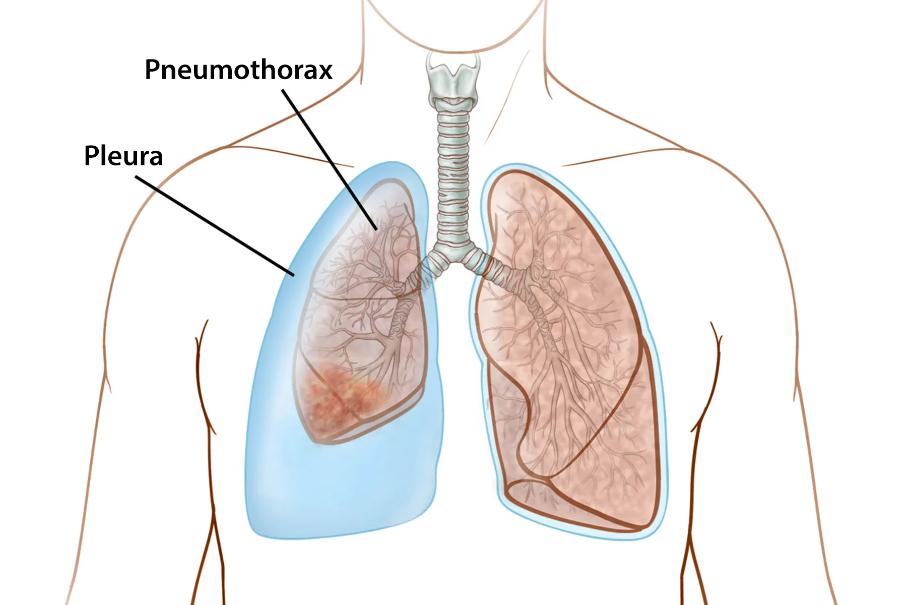
A pneumothorax, commonly called a collapsed lung, sounds like the kind of thing that should arrive with dramatic movie music. Sometimes it does. A person may feel sudden sharp chest pain, become short of breath, and realize very quickly that this is not a “walk it off” situation. Other times, especially when the collapse is small, the symptoms can be subtle enough to make someone wonder whether they pulled a muscle, slept weird, or angered their left rib cage.
In plain English, pneumothorax happens when air gets into the space between the lung and the chest wall. That space is called the pleural space. Your lung normally expands like a soft balloon when you breathe in, but trapped air outside the lung can press on it and keep it from fully expanding. The result may be a partial or complete lung collapse.
The good news is that pneumothorax is treatable. The very important news is that it can become an emergency, especially when pressure builds quickly or breathing becomes difficult. This guide explains pneumothorax symptoms, causes, diagnosis, treatment options, recovery, prevention, and real-life-style experiences that help make the condition easier to understand.
What Is Pneumothorax?
Pneumothorax is the medical term for air in the pleural space. The pleura is a thin lining around the lungs and inside the chest wall. Under normal conditions, this area helps the lungs glide smoothly as you breathe. When air leaks into that space, it can push against the lung and cause it to collapse partially or completely.
Think of your lung as a balloon inside a protective box. If air sneaks into the box but outside the balloon, the balloon gets squeezed. That is basically pneumothorax, minus the party decorations.
A collapsed lung can affect one side of the chest or, rarely, both sides. The severity depends on how much air is trapped, how quickly it accumulates, whether the person has underlying lung disease, and whether the pressure affects the heart and blood flow.
Common Symptoms of Pneumothorax
The classic symptoms of pneumothorax are sudden chest pain and shortness of breath. The chest pain is often sharp, one-sided, and worse when taking a deep breath or coughing. Some people describe it as stabbing, tight, or like something “popped” inside the chest.
Possible pneumothorax symptoms include:
- Sudden sharp chest pain, often on one side
- Shortness of breath or trouble taking a full breath
- Chest tightness or pressure
- Rapid heartbeat
- Fast or shallow breathing
- Fatigue or weakness
- Lightheadedness or feeling faint
- Bluish lips, skin, or fingernails from low oxygen
- Shoulder, back, or upper abdominal discomfort
- Anxiety or a sense that something is seriously wrong
Symptoms can vary. A small pneumothorax may feel like mild chest discomfort and slight breathlessness. A large pneumothorax can make breathing hard and frightening. A tension pneumothorax, the most dangerous form, can cause low blood pressure, severe breathing distress, confusion, shock, or collapse.
When Is a Collapsed Lung an Emergency?
Any sudden chest pain with shortness of breath deserves urgent medical attention. Do not try to diagnose pneumothorax at home with internet confidence and a mirror. The chest is not a DIY project.
Call emergency services or go to the nearest emergency department if you have:
- Severe or worsening shortness of breath
- Sharp chest pain that starts suddenly
- Blue lips, fingernails, or skin
- Fainting, confusion, or extreme weakness
- Chest symptoms after a car crash, fall, sports injury, stabbing, gunshot wound, or rib injury
- Rapid heartbeat with breathing difficulty
- Known lung disease and new chest pain or breathlessness
A tension pneumothorax is a life-threatening emergency. In this situation, air keeps entering the pleural space and cannot escape. Pressure builds in the chest, compressing the lung and potentially shifting structures in the chest. It can interfere with the heart’s ability to pump blood. Immediate medical treatment is required.
Types of Pneumothorax
Doctors classify pneumothorax by cause and severity. Understanding the type helps guide treatment.
Primary spontaneous pneumothorax
Primary spontaneous pneumothorax happens without an obvious injury or known lung disease. It is often linked to small air-filled blisters on the lung surface, called blebs, that rupture. It is more common in tall, thin young adults and people who smoke.
Secondary spontaneous pneumothorax
Secondary spontaneous pneumothorax occurs in people with existing lung disease. Conditions such as chronic obstructive pulmonary disease, asthma, cystic fibrosis, pneumonia, tuberculosis, lung cancer, or interstitial lung disease can weaken lung tissue and increase risk. Because the lungs may already be compromised, symptoms can be more severe even if the pneumothorax is not large.
Traumatic pneumothorax
Traumatic pneumothorax happens after chest injury. A broken rib, blunt trauma, penetrating wound, or serious accident can puncture the lung or allow air into the chest cavity. This type needs prompt evaluation because injuries may involve bleeding, rib fractures, or damage to other organs.
Iatrogenic pneumothorax
Iatrogenic pneumothorax is caused by a medical procedure. It can occur after procedures such as lung biopsy, central line placement, mechanical ventilation, thoracentesis, or certain surgeries. This does not mean the procedure was careless; the lung is delicate, and sometimes anatomy behaves like a drama queen.
Tension pneumothorax
Tension pneumothorax is the most dangerous type. Air builds under pressure and cannot escape. It can rapidly impair breathing and circulation. This is treated immediately, often before all imaging is complete, because waiting can be dangerous.
What Causes Pneumothorax?
Pneumothorax may occur for several reasons. Sometimes the cause is obvious, such as a chest injury. Other times, it appears suddenly in someone who seemed healthy.
Common causes and risk factors include:
- Smoking or vaping, which can damage lung tissue
- Underlying lung disease such as COPD, asthma, cystic fibrosis, or pneumonia
- Chest trauma from accidents, falls, or sports injuries
- Medical procedures involving the chest or lungs
- Mechanical ventilation, especially with high airway pressure
- Family history of spontaneous pneumothorax
- Previous pneumothorax
- Rapid pressure changes, such as scuba diving or high-altitude exposure
- Rare genetic or connective tissue disorders
Smoking is one of the most important preventable risk factors. It raises the chance of spontaneous pneumothorax and recurrence. Quitting smoking is not always easy, but neither is having a chest tube, so the motivation is fairly strong.
How Doctors Diagnose Pneumothorax
Diagnosis usually starts with symptoms, medical history, and a physical exam. A clinician may listen to the lungs with a stethoscope and notice reduced or absent breath sounds on the affected side. They may also check oxygen levels, heart rate, breathing rate, and blood pressure.
Tests may include:
- Chest X-ray: Often the first imaging test used to confirm air outside the lung.
- Chest CT scan: Provides more detailed images and may detect small pneumothoraces or underlying lung problems.
- Ultrasound: Often useful in emergency and trauma settings because it can be done quickly at the bedside.
- Arterial blood gas test: Measures oxygen and carbon dioxide levels in the blood when breathing problems are significant.
- Pulse oximetry: A small finger sensor checks blood oxygen saturation.
Doctors also determine whether the pneumothorax is small or large, stable or worsening, simple or tension-related, and whether another condition caused it. Treatment depends on these details.
Pneumothorax Treatment Options
The main goal of pneumothorax treatment is simple: remove air from the pleural space, help the lung re-expand, prevent complications, and reduce the risk of recurrence. The exact treatment depends on the size of the pneumothorax, symptoms, cause, and overall health.
1. Observation
A small, uncomplicated pneumothorax may heal on its own. In these cases, doctors may monitor the patient, repeat chest imaging, and provide instructions for follow-up care. The body can slowly absorb the trapped air, allowing the lung to re-expand.
Observation is only appropriate when the person is stable and symptoms are mild. It is not a green light to ignore worsening chest pain or shortness of breath.
2. Supplemental oxygen
Oxygen therapy may help improve oxygen levels and can support the body while it absorbs air from the pleural space. It may be used during observation or alongside other treatments.
3. Needle aspiration
Needle aspiration involves inserting a needle or small catheter into the chest to remove trapped air. This may be used for some larger or symptomatic pneumothoraces. It can relieve pressure and allow the lung to re-expand.
4. Chest tube insertion
A chest tube, also called tube thoracostomy, is a hollow tube placed between the ribs into the pleural space. It drains air and sometimes fluid from around the lung. The tube may be connected to suction or a drainage device. It stays in place until the air leak improves and the lung remains expanded.
Chest tubes sound intimidating, and honestly, nobody adds one to their vacation wish list. But they can be lifesaving and highly effective when air needs to be removed continuously.
5. Emergency needle decompression
For tension pneumothorax, emergency needle decompression may be performed immediately to release trapped air and reduce pressure. A chest tube is typically placed afterward for continued drainage.
6. Surgery
Surgery may be recommended for recurrent pneumothorax, persistent air leak, bilateral pneumothorax, certain high-risk jobs or activities, or cases that do not respond to less invasive treatment. Many procedures are done with video-assisted thoracoscopic surgery, known as VATS. Surgeons may remove blebs and perform pleurodesis, a technique that helps the lung stick to the chest wall to reduce recurrence risk.
Recovery After Pneumothorax
Recovery time varies widely. A small pneumothorax may improve over days to a few weeks. A larger pneumothorax requiring a chest tube or surgery may need a longer recovery period. People with underlying lung disease may take more time to return to normal activities.
Follow-up is important. Doctors often repeat chest imaging to confirm that the lung has re-expanded and that air is not returning. Even when someone feels better, the inside of the chest may still be finishing its repair work. The body is talented, but it does not always send calendar invites.
During recovery, patients may be told to:
- Avoid smoking and secondhand smoke
- Rest and gradually return to activity
- Avoid heavy lifting until cleared
- Keep follow-up appointments
- Watch for returning chest pain or shortness of breath
- Avoid flying, scuba diving, high-altitude travel, or playing wind instruments until a clinician says it is safe
- Take prescribed pain medicine as directed
- Care for any incision or chest tube site according to instructions
Anyone who has had pneumothorax should ask their clinician about activity restrictions. Scuba diving is especially risky after spontaneous pneumothorax unless a specialist provides specific clearance after definitive treatment.
Can Pneumothorax Come Back?
Yes, pneumothorax can recur. The risk is higher in people who have had one before, people who smoke, and people with underlying lung disease. Recurrence risk is one reason follow-up care matters so much.
Prevention depends on the cause. Not every pneumothorax can be prevented, but several steps can reduce risk:
- Quit smoking and avoid vaping
- Manage chronic lung conditions carefully
- Use protective gear during high-risk sports or work
- Follow medical advice after chest procedures
- Avoid unsafe pressure-change activities after pneumothorax
- Report new chest symptoms early
Pneumothorax vs. Atelectasis: Are They the Same?
Pneumothorax and atelectasis can both involve lung collapse, but they are not the same thing. Pneumothorax is caused by air in the pleural space pressing on the lung from the outside. Atelectasis usually happens when air sacs inside the lung collapse, often because of blockage, shallow breathing after surgery, mucus plugs, or pressure from outside the lung.
Both can cause shortness of breath, but they have different causes and treatments. This is another reason chest imaging and medical evaluation are essential.
Living With a History of Pneumothorax
After a collapsed lung, many people feel cautious about every chest sensation. That is understandable. A random twinge near the ribs can suddenly feel like breaking news. The key is to know your warning signs, follow your care plan, and ask your healthcare provider what is normal during recovery.
It can also help to keep a simple record of symptoms, activity levels, follow-up visits, and restrictions. If chest pain returns, especially with shortness of breath, do not try to “wait and see” for too long. Pneumothorax is much easier to treat when it is recognized early.
Practical Experiences and Real-Life Lessons About Pneumothorax
Because pneumothorax can appear suddenly, the experience often teaches people a few memorable lessons. These examples are not individual medical advice, but they reflect common situations people may encounter.
The “I thought it was a pulled muscle” experience
Some people first notice sharp pain on one side of the chest after coughing, exercising, or simply standing up. They may assume they strained a muscle. That is understandable because chest wall pain can feel sharp too. The difference is that pneumothorax pain often comes with shortness of breath, difficulty taking a deep breath, or a strange tight feeling inside the chest. If breathing feels different, the safest move is medical evaluation. Your lung does not appreciate being ignored like an unread email.
The “healthy person surprise” experience
Primary spontaneous pneumothorax can happen to people who do not think of themselves as sick. A tall, thin young adult may suddenly feel chest pain while resting, walking, or doing routine activity. This can be confusing because there may be no accident and no obvious trigger. The lesson: looking healthy does not rule out pneumothorax. Sudden chest pain plus breathing trouble is always worth taking seriously.
The “chest tube was scary but helped” experience
For larger pneumothorax, a chest tube may be needed. Many patients feel nervous when they hear those words. That reaction is normal. A tube in the chest sounds like something invented by a committee with no interest in comfort. But the purpose is practical: it removes air so the lung can expand again. People often feel breathing improve once pressure is relieved. There may be soreness at the tube site, but nurses and doctors monitor pain, drainage, air leaks, and lung expansion carefully.
The “recovery is not a race” experience
After discharge, a person may feel better but still tire easily. Walking across the room may be fine, while stairs feel like a dramatic mountain expedition. Recovery often requires patience. The lung needs time to stay expanded and heal. Returning to heavy exercise, flying, diving, or lifting too soon may create risk. The best approach is to follow the clinician’s timeline, not the timeline suggested by boredom, optimism, or a gym membership renewal notice.
The “stop smoking became real” experience
Many people hear for years that smoking affects the lungs. A pneumothorax can make that warning feel very personal. Smoking increases the risk of spontaneous pneumothorax and can contribute to recurrence. For someone who has already had a collapsed lung, quitting is one of the most meaningful prevention steps. Support may include counseling, nicotine replacement, prescription medications, quitlines, and help from a primary care clinician. Quitting is difficult, but breathing is a persuasive hobby.
The “follow-up appointment mattered” experience
Once symptoms improve, it is tempting to skip follow-up. However, imaging may be needed to confirm the lung remains expanded. A person may feel nearly normal while a small amount of air is still present. Follow-up visits also help clarify when it is safe to work, exercise, travel, or resume specific activities. This is especially important for people whose jobs involve flying, diving, remote travel, heavy labor, or limited access to emergency care.
The “anxiety after pneumothorax is normal” experience
A collapsed lung can be frightening. Afterward, people may feel anxious about breathing, sleeping positions, chest sensations, or being far from a hospital. This worry usually improves as recovery progresses and follow-up tests look good. Still, emotional recovery deserves attention too. Ask questions, request clear instructions, and talk with a healthcare provider if fear interferes with daily life. Peace of mind is part of healing, not a luxury add-on.
Conclusion
Pneumothorax, or collapsed lung, happens when air collects between the lung and chest wall, pressing on the lung and preventing normal expansion. The most common symptoms are sudden chest pain and shortness of breath, but signs can range from mild discomfort to life-threatening distress. Diagnosis usually involves a physical exam and imaging such as a chest X-ray, CT scan, or ultrasound.
Treatment depends on severity. Small, stable cases may only need observation and oxygen. Larger or more serious cases may require needle aspiration, chest tube drainage, emergency decompression, or surgery. Recovery involves follow-up imaging, activity restrictions, lung care, and prevention steps such as quitting smoking and managing existing lung disease.
The big takeaway is simple: do not ignore sudden chest pain with breathing trouble. Pneumothorax is treatable, but timing matters. When your lungs ask for help, answer the call quickly.