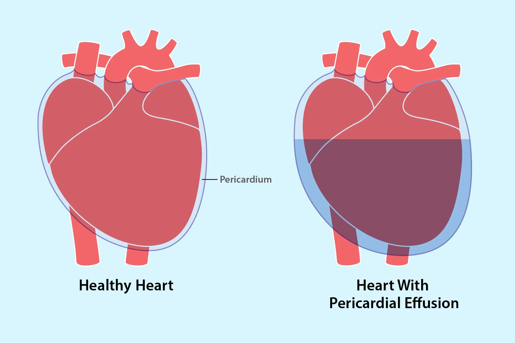
Your heart is many things: hardworking, dramatic, and absolutely not a fan of cramped living conditions. Pericardial effusion happens when extra fluid collects in the sac around the heart, called the pericardium. A tiny amount of fluid there is normal. Too much is not. When the buildup becomes significant, it can squeeze the heart, limit how well it fills, and turn a medical problem into a medical emergency.
That is why pericardial effusion matters. Some cases are small, quiet, and found by accident during imaging. Others announce themselves with chest pressure, shortness of breath, dizziness, or a racing heartbeat. The biggest concern is cardiac tamponade, a dangerous situation in which the pressure from fluid prevents the heart from pumping effectively.
This guide explains what pericardial effusion is, what causes it, what symptoms to watch for, how doctors diagnose it, and how treatment works. It is written in plain English, with enough depth to be useful and enough humanity to keep it from reading like a textbook that skipped its coffee.
Important note: This article is for education only and is not a substitute for medical care. Chest pain, fainting, severe shortness of breath, or symptoms of shock need urgent evaluation.
What Is Pericardial Effusion?
The heart sits inside a thin, two-layered sac called the pericardium. Normally, there is a small amount of lubricating fluid between those layers so the heart can beat without friction. Pericardial effusion means there is too much fluid in that space.
Here is the tricky part: the danger is not just about how much fluid is present. It is also about how fast the fluid builds up. If it accumulates slowly, the pericardium may stretch enough to make room for quite a bit of fluid before symptoms become obvious. If it builds up quickly, even a smaller amount can put sudden pressure on the heart and cause major problems.
In other words, pericardial effusion is a bit like traffic in a tunnel. A few extra cars may not matter if they arrive gradually. A sudden pileup, however, changes the whole commute. Fast.
What Causes Pericardial Effusion?
Pericardial effusion is not one disease with one simple cause. It is more like a sign that something is irritating, inflaming, injuring, or otherwise affecting the pericardium.
1. Pericarditis and Other Inflammatory Conditions
One of the most common pathways is pericarditis, which is inflammation of the pericardium. When the sac becomes inflamed, fluid can increase. This may happen after a viral illness, after a heart attack, or after heart surgery or other heart procedures.
2. Infections
Infections can trigger fluid buildup around the heart. Viral infections are common culprits, but bacterial infections and other less common infections can also be involved. When infection is suspected, doctors may analyze the pericardial fluid to help identify the cause.
3. Autoimmune and Inflammatory Diseases
Conditions such as lupus and rheumatoid arthritis can inflame the pericardium and lead to effusion. In these cases, the immune system is essentially misfiring and stirring up inflammation where it is not wanted.
4. Cancer
Cancer may cause pericardial effusion either by involving the pericardium directly or by spreading from another site. Lung cancer, breast cancer, lymphoma, and other malignancies may be associated with fluid around the heart.
5. Kidney Failure and Metabolic Causes
People with advanced kidney failure can develop pericardial effusion because waste products build up in the blood and irritate the pericardium. This is sometimes referred to as uremic pericardial disease.
6. Hypothyroidism
An underactive thyroid can also be linked with fluid accumulation around the heart. It is not the first cause most people guess, but the thyroid has a habit of quietly involving itself in places you did not invite it.
7. Injury, Trauma, and Medical Procedures
Chest trauma, bleeding after an injury, and complications from medical procedures involving the heart can all cause fluid or blood to collect in the pericardial space.
8. Radiation, Medicines, and Unknown Causes
Radiation therapy near the chest and certain medications can contribute to pericardial effusion. And sometimes, even after a thorough workup, no clear cause is found. That can be frustrating, but it is a real part of clinical practice.
Pericardial Effusion Symptoms: What Does It Feel Like?
Symptoms vary widely. Some people have no symptoms at all, especially if the effusion is small or developed slowly. Others feel distinctly unwell.
Common Symptoms
- Shortness of breath
- Chest pain, pressure, or discomfort
- Difficulty breathing when lying flat
- Fast heartbeat or palpitations
- Lightheadedness or dizziness
- Fainting
- Fatigue
- Swelling in the legs, belly, or sometimes the arms
Some people may also notice cough, hoarseness, hiccups, nausea, or a sense of fullness in the chest if a large effusion presses on nearby structures.
Emergency Warning Signs
If the fluid begins to compress the heart, symptoms can become more dramatic. Red flags include:
- Chest pain that lasts more than a few minutes
- Severe shortness of breath or painful breathing
- Unexplained fainting
- Very low blood pressure
- Cool or clammy skin
- Rapid breathing
- Confusion or extreme weakness
These may signal cardiac tamponade or shock, both of which require emergency care.
How Pericardial Effusion Is Diagnosed
Diagnosis starts with the basics: symptoms, medical history, and a physical exam. A doctor will ask when symptoms began, whether they are getting worse, and whether there are possible clues such as recent infection, autoimmune disease, cancer history, kidney disease, heart surgery, or chest injury.
Echocardiogram: The Star Player
The most important test is usually an echocardiogram, which is an ultrasound of the heart. It can show how much fluid is present and whether the heart is being compressed. It also helps determine whether the heart’s chambers are having trouble filling properly.
Other Imaging Tests
- Electrocardiogram (ECG/EKG): Looks at the heart’s electrical activity and may show patterns that raise concern for tamponade or related heart strain.
- Chest X-ray: Can sometimes show an enlarged heart silhouette when the effusion is large.
- CT or MRI: May reveal fluid around the heart and provide added detail, especially when imaging is being done for another reason or when the anatomy needs closer review.
Blood Tests and Fluid Analysis
Blood tests may help doctors look for the underlying cause. If fluid is drained, that sample can be studied for infection, cancer cells, inflammation, and other clues. In uncertain cases, this step can be just as valuable as removing the fluid itself.
Pericardial Effusion Treatment
Treatment depends on three main things: the amount of fluid, the cause, and whether tamponade is present or likely.
1. Monitoring and Treating the Underlying Cause
Not every pericardial effusion needs a dramatic intervention. Small effusions may be monitored with follow-up imaging while doctors treat the condition behind it. For example, if inflammation is driving the problem, controlling the inflammation may allow the fluid to improve.
2. Medications
When the effusion is related to inflammation and there is no immediate tamponade, medications may be used. These can include:
- Aspirin
- NSAIDs such as ibuprofen
- Colchicine
- Corticosteroids such as prednisone in selected cases
The exact choice depends on the suspected cause and the person’s other medical issues. For example, a doctor may avoid certain anti-inflammatory medicines in someone with kidney disease or other risk factors.
3. Pericardiocentesis
Pericardiocentesis is a procedure that uses a needle and usually a catheter to drain fluid from the pericardial sac. Imaging guidance, often echocardiography, helps position the needle safely. This can relieve pressure on the heart and also provide fluid for lab testing.
Pericardiocentesis may be lifesaving when tamponade is present. It is also often used when the cause of the effusion is unclear, when symptoms are significant, or when the fluid keeps returning.
4. Surgical Drainage or a Pericardial Window
Some patients need more than needle drainage. A pericardial window is a surgical procedure in which a small part of the pericardium is removed so fluid can drain more continuously. This may be recommended if the fluid comes back, if the anatomy makes pericardiocentesis difficult, or if tissue needs to be examined.
In some cases, more extensive surgery may be needed, especially when bleeding is involved or when the effusion is recurrent and stubborn.
5. Pericardiectomy in Recurrent Cases
If the effusion repeatedly returns after drainage, doctors may consider removing part or all of the pericardium. This is called pericardiectomy. It is not the first step for most people, but it can become the right step in selected cases.
Possible Complications
The complication everyone worries about most is cardiac tamponade. This happens when the pressure around the heart becomes high enough to prevent the chambers from filling normally. Less blood then leaves the heart, which means less oxygen reaches the body.
If untreated, tamponade can lead to shock, organ failure, and death. Rarely, longstanding inflammation can also lead to scarring or stiffening of the pericardium, making it harder for the heart to function normally over time.
When to Seek Urgent Medical Care
Call emergency services or seek immediate medical attention if you have:
- Chest pain that is severe or lasts more than a few minutes
- Trouble breathing
- Fainting or nearly fainting
- Rapid worsening of weakness, dizziness, or confusion
- Symptoms of shock, such as clammy skin, severe shortness of breath, or a feeling that you may pass out
Pericardial effusion can sometimes be monitored safely, but it is not a condition to self-diagnose with confidence after five minutes on the internet and one dramatic sigh. When symptoms are serious, evaluation needs to be prompt.
Questions to Ask Your Doctor
- What is the most likely cause of my pericardial effusion?
- How large is it, and is it affecting how my heart works?
- Do I need only monitoring, or do I need drainage?
- What tests can help find the cause?
- What symptoms should make me go to the ER right away?
- What is the chance this will come back?
- Do I need follow-up echocardiograms?
Common Experiences Related to Pericardial Effusion
One of the most overlooked parts of pericardial effusion is what the experience feels like from the patient side. The diagnosis often arrives with a strange mix of confusion and fear. Many people do not wake up thinking, “Today I will learn there is fluid around my heart.” More often, they go in for chest discomfort, shortness of breath, fatigue, or dizziness and expect to hear something mundane. Then a scan, an ultrasound, or an echocardiogram changes the entire tone of the day.
For some patients, the emotional whiplash is the hardest part at first. Symptoms may have been mild, vague, or easy to dismiss. A little breathlessness while climbing stairs. A little chest pressure. A little extra fatigue. Nothing that screams “heart sac problem” to the average human trying to get through the week. That is why many people feel stunned when they hear the diagnosis. The term sounds serious because it is serious, but the next question is always the same: How serious is mine?
Another common experience is uncertainty while doctors search for the cause. A patient may hear that the effusion could be linked to inflammation, infection, autoimmune disease, cancer, kidney problems, prior surgery, or sometimes no obvious cause at all. That uncertainty can be exhausting. It is one thing to know fluid is present. It is another thing entirely to wait for blood work, imaging, or fluid analysis to explain why it is there.
People who need drainage often describe a combination of relief and anxiety. Relief, because they finally have a plan. Anxiety, because anything involving a needle near the heart sounds like a sentence invented by a screenwriter who wanted to raise the stakes. In reality, procedures such as pericardiocentesis are done with imaging guidance and careful monitoring, but it is completely normal for patients to feel nervous beforehand.
Recovery brings its own emotions. Some people feel better quickly, especially if the fluid was causing significant pressure. Breathing becomes easier. Chest pressure lifts. Energy starts to come back. Others recover more gradually, especially when the effusion is part of a larger illness. Follow-up appointments, repeat echocardiograms, medication adjustments, and the possibility of recurrence can make people feel as if they are living from one checkpoint to the next.
Families go through a parallel experience. They are often the ones asking practical questions, taking notes, remembering medication names, and pretending to be calmer than they feel. If the word “tamponade” enters the conversation, stress levels rise fast. Good communication from the healthcare team matters enormously here. When patients and loved ones understand the plan, what symptoms to watch for, and what follow-up is needed, the situation becomes less mysterious and more manageable.
Perhaps the most reassuring shared experience is this: many people do improve, especially when the cause is identified and treated appropriately. The road can be bumpy, but it is not automatically hopeless. Knowledge, prompt evaluation, and good follow-up make a real difference.
Final Thoughts
Pericardial effusion is a condition that ranges from mild and manageable to urgent and life-threatening. The difference often comes down to the amount of fluid, the speed of buildup, the underlying cause, and whether the heart is being compressed.
The good news is that modern diagnosis and treatment are highly effective. Echocardiography helps doctors see what is happening in real time. Medications can calm inflammation. Drainage procedures can relieve pressure and provide answers. Surgery is available when recurring or complex cases demand a stronger fix.
The key is timing. Do not ignore persistent chest pain, breathing trouble, fainting, or sudden worsening symptoms. Your heart does excellent work every day. Giving it enough room to do that job is not optional.