Red eye can be dramatic. Sometimes it is allergies, dry eye, or the tragic aftermath of staring at spreadsheets like they personally offended you. But sometimes a red, painful, light-sensitive eye is something more serious: uveitis, a type of inflammation inside the eye that needs prompt medical attention.
Uveitis is not something your eye doctor diagnoses by squinting from across the room and saying, “Yep, looks angry.” The process is much more careful. A proper uveitis diagnosis usually combines your symptoms, medical history, a detailed eye exam, magnified inspection with a slit lamp, a dilated retinal exam, eye-pressure testing, and sometimes imaging or lab work. In other words, your eye doctor becomes part detective, part photographer, part microscope wizard.
This guide explains how an ophthalmologist or optometrist figures out whether you have uveitis, what each test is looking for, and why your eye doctor may ask questions that seem to have nothing to do with your eyeballslike joint pain, back stiffness, rashes, infections, or autoimmune disease history. Spoiler: the eye is connected to the rest of the body, even when it acts like an independent drama department.
What Is Uveitis?
Uveitis means inflammation inside the eye, often involving the uvea, the middle layer of the eye that includes the iris, ciliary body, and choroid. However, the term can also include inflammation affecting nearby structures such as the retina, vitreous, or optic nerve. That is why uveitis can look different from person to person.
Doctors usually classify uveitis by where the inflammation appears:
Anterior Uveitis
This is the most common type and affects the front part of the eye, especially the iris. It may also be called iritis. Symptoms often include redness, pain, light sensitivity, blurry vision, and sometimes a smaller or irregular pupil.
Intermediate Uveitis
This affects the middle area of the eye, including the vitreous gel. Floaters and blurred vision may be more noticeable than redness or pain.
Posterior Uveitis
This affects the back of the eye, including the retina or choroid. It may cause floaters, blurry vision, blind spots, or reduced vision without much pain. Sneaky? Absolutely.
Panuveitis
This means inflammation involves multiple areas of the eye: front, middle, and back. It usually requires a more detailed evaluation and close follow-up.
When Your Symptoms Raise Suspicion
Your eye doctor starts by listening. That may sound old-fashioned in a world full of machines, scans, and blinking lights, but symptoms provide major clues. Uveitis often announces itself with one or more of the following:
- Eye redness, especially around the colored part of the eye
- Eye pain or aching
- Sensitivity to light
- Blurred vision
- Floaters, spots, or cobweb-like shadows
- Reduced vision
- Tearing or discomfort
- A pupil that looks small or oddly shaped
A red eye with pain and light sensitivity is especially important. Conjunctivitis can make your eye red and goopy, but uveitis often causes deeper aching and a strong “turn off the sun immediately” reaction. If vision changes are present, your eye doctor will take the situation even more seriously.
The Medical History: The Detective Work Begins
Before the exam gets technical, your doctor will ask questions. Some may feel surprisingly unrelated. Do you have back pain? Joint stiffness? Mouth ulcers? Skin rashes? Digestive problems? Recent infections? A history of shingles, tuberculosis exposure, syphilis, Lyme disease, inflammatory bowel disease, sarcoidosis, ankylosing spondylitis, juvenile idiopathic arthritis, or autoimmune disease?
These questions matter because uveitis can be idiopathic, meaning no clear cause is found, but it can also be linked to infections, autoimmune conditions, inflammatory disorders, eye injuries, or previous eye surgery. Your eye may be the first body part to wave a tiny red flag.
Your doctor may ask whether this is your first episode or whether it keeps coming back. A single mild episode of anterior uveitis may be handled differently from recurrent, bilateral, severe, or unusual inflammation. Recurrent uveitis can push the doctor to look harder for an underlying systemic condition.
Visual Acuity Test: How Well Are You Seeing?
The first hands-on step is usually checking your vision. You may read letters on a chart, identify shapes, or use a pinhole device if you do not have your glasses with you. This test may look basic, but it gives the doctor a starting point.
Visual acuity helps answer several key questions: Has inflammation affected central vision? Is one eye worse than the other? Is vision loss mild, moderate, or urgent? If your vision is significantly reduced, the doctor may look more carefully for swelling in the retina, inflammation in the vitreous, optic nerve involvement, or complications such as glaucoma or cataract.
Think of this test as the “before photo” in the uveitis story. It helps your doctor measure whether treatment is working later.
External Eye Exam: Looking for Surface Clues
Next, your doctor examines the outside of the eye. They look at the lids, lashes, conjunctiva, cornea, pupil shape, and the pattern of redness. Uveitis often causes redness near the cornea, sometimes called ciliary flush or limbal injection. That pattern can look different from the more widespread redness seen with simple conjunctivitis.
The doctor may also check how your pupils react to light. If the iris is inflamed, the pupil may be small, sluggish, or irregular. In some cases, inflammation can cause the iris to stick to the lens, creating a condition called posterior synechiae. That sounds like a villain from a sci-fi movie, but it is really scar tissue that can affect pupil shape and eye function.
Slit-Lamp Exam: The Star of Uveitis Diagnosis
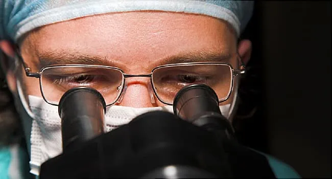
If uveitis diagnosis had a main character, it would be the slit-lamp exam. A slit lamp is a microscope with a bright, narrow beam of light that lets your eye doctor examine the front part of your eye in high detail.
During this exam, you sit with your chin and forehead against supports while the doctor shines a thin light into your eye. It may feel bright, but it should not be painful. Your job is simple: hold still and resist the heroic urge to blink at exactly the wrong moment.
The slit lamp helps your doctor look for tiny inflammatory cells floating in the anterior chamber, the fluid-filled space between the cornea and iris. These white blood cells are one of the classic signs of anterior uveitis. The doctor may also see “flare,” which means protein leakage into the eye fluid, making it look hazy under magnification.
Other slit-lamp clues may include keratic precipitates, which are clusters of inflammatory cells on the back surface of the cornea; hypopyon, a visible layer of white blood cells in severe inflammation; iris nodules; fibrin; or adhesions between the iris and lens. To the patient, it may look like the doctor is just peering intensely. To the doctor, your eye is basically giving a PowerPoint presentation titled “Inflammation: A Memoir.”
Eye Pressure Test: Tonometry
Uveitis can affect eye pressure. Sometimes inflammation lowers intraocular pressure. Other times, it raises pressure, especially if inflammatory debris blocks fluid drainage or if steroid treatment later increases pressure. That is why your eye doctor often performs tonometry, a test that measures pressure inside the eye.
There are several ways to measure eye pressure. Some devices gently touch the numbed eye surface, while others use a quick puff of air. No, the air puff is not your eye doctor’s revenge for you missing last year’s appointment, though it can feel personal.
Pressure matters because high eye pressure can damage the optic nerve and contribute to glaucoma. When uveitis is present, tracking pressure is part of protecting long-term vision.
Dilated Eye Exam: Checking the Back of the Eye
After examining the front of the eye, your doctor may use dilating drops to widen your pupils. Dilation allows a better view of the lens, vitreous, retina, choroid, blood vessels, and optic nerve.
This step is important because not all uveitis stays in the front of the eye. Intermediate, posterior, and panuveitis may involve inflammation deeper inside the eye. The doctor may look for vitreous cells, retinal swelling, inflamed blood vessels, choroidal lesions, optic nerve swelling, or scars from previous inflammation.
Dilation can make your vision blurry and your eyes sensitive to light for several hours. Bring sunglasses. If you forget them, you may leave the office doing the classic “vampire exiting a movie theater at noon” squint.
OCT Imaging: A Cross-Section of the Retina
Optical coherence tomography, or OCT, is a noninvasive imaging test that creates detailed cross-sectional pictures of the retina and sometimes other eye structures. It is fast, painless, and useful when the doctor needs to check for swelling, fluid, or structural changes.
In uveitis, OCT can help detect macular edema, which is swelling in the central retina. This matters because the macula is responsible for sharp central vision. Even if the front of the eye looks calmer, hidden retinal swelling can still affect reading, driving, and recognizing faces.
OCT can also help monitor whether treatment is working. If swelling decreases over time, the scan provides objective evidence that inflammation is improving. Your retina does not get to say, “I’m fine,” while secretly hoarding fluid.
Fundus Photography: Documenting the Scene
Your doctor may take color photographs of the back of your eye. These images document what the retina, optic nerve, and blood vessels look like at the time of diagnosis. They are useful for comparison during follow-up visits.
Fundus photography may capture scars, retinal lesions, blood vessel inflammation, optic nerve changes, or other findings. In uveitis care, comparison over time is powerful. A photo from last month may reveal whether a spot has faded, grown, or stayed stable.
Fluorescein Angiography and Other Dye Tests
If your doctor suspects retinal blood vessel leakage, inflammation, or circulation problems, they may order fluorescein angiography. In this test, a dye is injected into a vein in your arm, and special photographs are taken as the dye travels through the blood vessels in the eye.
This test can show leakage from inflamed vessels, swelling near the optic nerve, areas of poor blood flow, or retinal vasculitis. Another dye test, indocyanine green angiography, may be used when the doctor needs more information about the choroid, the vascular layer beneath the retina.
Dye tests sound intense, but they are common in retinal and inflammatory eye disease evaluation. You may notice temporary yellowish skin or bright yellow urine afterward. This is usually expected, not a sign that you are turning into a highlighter.
When Lab Tests Are Needed
Not every person with uveitis needs a giant lab workup. In fact, smart uveitis evaluation is targeted. Eye doctors usually decide on testing based on the pattern of inflammation, whether one or both eyes are involved, whether the condition is recurrent, your age, symptoms outside the eye, travel history, infection risks, and exam findings.
Possible tests may include blood work for inflammatory markers, syphilis testing, tuberculosis screening, HLA-B27 testing, tests related to sarcoidosis, Lyme disease testing in appropriate regions or exposure situations, and other autoimmune or infectious evaluations. A chest X-ray or chest imaging may be considered if sarcoidosis or tuberculosis is suspected. In certain cases, your doctor may coordinate with a primary care physician, rheumatologist, infectious disease specialist, or neurologist.
The goal is not to order every test known to modern civilization. The goal is to ask, “What causes fit this patient’s exact pattern?” That approach prevents unnecessary confusion, false positives, and medical bills that arrive wearing tap shoes.
Eye Fluid Testing: Rare, But Sometimes Important
In unusual, severe, persistent, or infection-suspected cases, the doctor may consider testing fluid from inside the eye, such as aqueous or vitreous fluid. This is not part of a routine exam for most patients. It is generally reserved for situations where the diagnosis is unclear or where identifying an infectious cause could change treatment.
For example, if viral, fungal, parasitic, or masquerade conditions are being considered, fluid analysis may provide information that cannot be obtained from a standard eye exam alone. This is one reason complex uveitis is often managed by or referred to an ophthalmologist with experience in ocular inflammation.
Ruling Out Look-Alike Conditions
Uveitis can mimic or overlap with other eye problems. Your doctor may consider conditions such as conjunctivitis, keratitis, scleritis, acute angle-closure glaucoma, corneal abrasion, dry eye, retinal tear, infection, or trauma. This is why self-diagnosing a painful red eye is risky.
For example, conjunctivitis is often itchy or discharge-heavy. Keratitis may involve corneal staining and severe surface pain. Acute angle-closure glaucoma can cause high eye pressure, headache, nausea, halos, and a dangerously firm eye. Uveitis has its own clues, but the exam is what separates “annoying” from “urgent.”
How Doctors Determine the Type and Severity
Once your doctor sees inflammation, they classify it. They may describe the location, severity, duration, and pattern:
- Location: anterior, intermediate, posterior, or panuveitis
- Onset: sudden or gradual
- Duration: limited, persistent, or chronic
- Course: first episode, recurrent, or continuous
- Laterality: one eye or both eyes
- Appearance: granulomatous or non-granulomatous inflammation
- Complications: macular edema, glaucoma, cataract, synechiae, retinal damage, or optic nerve involvement
This classification helps guide treatment and follow-up. Mild anterior uveitis may need one plan; posterior uveitis with retinal vascular leakage may need a very different one. Same umbrella term, different weather forecast.
What Happens After Diagnosis?
Although this article focuses on how your eye doctor figures out if you have uveitis, diagnosis and treatment usually happen close together. Uveitis can threaten vision, so doctors often treat inflammation promptly while continuing to investigate the cause.
Treatment may include corticosteroid eye drops, dilating drops to reduce pain and prevent iris adhesions, pressure-lowering drops if eye pressure is high, injections, oral medications, antimicrobial treatment if infection is suspected, or immune-modulating medicine for chronic noninfectious disease. Your exact plan depends on the type, severity, cause, and complications.
Follow-up is not optional decoration. Your doctor needs to confirm that inflammation is decreasing, pressure remains safe, and no hidden swelling or complications are developing. Uveitis can improve, flare, return, or change, which is why patients often need repeat exams.
When to Seek Urgent Eye Care
Do not wait days to “see if it clears up” if you have eye pain, light sensitivity, sudden floaters, blurred vision, vision loss, or a red eye that feels deeper than ordinary irritation. Same-day or urgent eye evaluation is wise, especially if symptoms are new, severe, or one-sided.
Uveitis is treatable, but delays can increase the risk of complications. Your future self, the one who enjoys seeing clearly, will appreciate your present self making the appointment.
Experience Section: What the Uveitis Exam May Feel Like in Real Life
For many patients, the uveitis evaluation starts with confusion. One day the eye is a little red. Then light feels rude. Then the computer screen looks foggy. By the time someone sits in the exam chair, they may be wondering whether they scratched the eye, caught an infection, or angered the contact lens gods.
The first surprise is often how many questions the doctor asks before touching any equipment. Patients sometimes expect a quick “look and prescribe drops” visit. Instead, the doctor may ask about back pain, arthritis, stomach issues, skin conditions, mouth sores, fevers, recent travel, sexually transmitted infection risks, tick bites, immune problems, and old eye injuries. It can feel like the appointment accidentally became a full-body interview. But that is exactly the point. Uveitis can be an eye problem with a body-wide backstory.
The slit-lamp exam is usually the moment when things become more specific. The patient sees a bright beam. The doctor sees cells, flare, corneal deposits, iris changes, or other microscopic details. A patient may hear the doctor say, “I see inflammation inside the eye.” That sentence can be unsettling, but it is also useful. It means the problem has moved from vague discomfort to a diagnosable pattern.
Dilation can be another memorable part of the experience. The drops may sting briefly, and after the pupils widen, reading a phone can become temporarily ridiculous. Many patients discover that parking-lot sunlight after dilation has the emotional intensity of a movie explosion. Sunglasses help. So does arranging a ride if vision is very blurry or if both eyes are dilated.
Imaging tests are usually easier than patients expect. OCT feels a bit like looking into a camera while trying not to blink. Fundus photos may flash brightly. Angiography, if needed, involves dye through a vein and a series of images. The idea of dye can make people nervous, but the test gives doctors important information about leakage, swelling, and blood vessel inflammation that may not be obvious from symptoms alone.
The most frustrating part of uveitis is that the first visit may not answer every “why.” Sometimes the doctor can confirm uveitis quickly but needs time to determine the cause. Sometimes no cause is ever found. That does not mean the exam failed. It means uveitis is a condition where pattern recognition, follow-up, and targeted testing matter more than one magical blood test.
A good patient experience often comes down to preparation. Bring a list of medications, medical conditions, recent infections, surgeries, autoimmune diagnoses, and family history. Mention symptoms outside the eye, even if they seem unrelated. Tell the doctor if this has happened before. Ask what warning signs should prompt urgent follow-up. And if drops are prescribed, ask exactly how often to use them and whether they should be shaken first. Eye drops love being misunderstood.
Most importantly, take follow-up seriously. Feeling better does not always mean inflammation is fully gone. Your doctor may need to taper medications carefully and check pressure, retina status, and signs of recurrence. With uveitis, the goal is not just to calm the red eye; it is to protect vision for the long run.
Conclusion
Figuring out whether you have uveitis is a careful process, not a guess. Your eye doctor combines your symptoms, medical history, visual acuity, slit-lamp findings, eye pressure, dilation, retinal examination, imaging, and selective lab tests to identify inflammation and understand its possible cause.
The key message is simple: painful red eye, light sensitivity, floaters, or blurred vision deserve prompt attention. Uveitis can be managed, but it needs the right diagnosis and follow-up. Your eye doctor has the tools to find the inflammation, classify it, monitor it, and protect your sight. Your job is to show up early, answer questions honestly, and not pretend the angry eye is “probably nothing” while it is clearly auditioning for a medical drama.